
Learn when you truly need an MRI scan or other imaging test to manage low back pain effectively.
See why it is so important to spot red flags and use your medical history to figure out what is causing your back pain.
Learn about other ways you can deal with your lower back that do not need imaging tests, and see why simple treatments can work well for problems with your lumbar spine.
Examine the primary causes of back pain and understand its widespread impact on people across India.
Understand the limits and possible risks of using MRI scans, CT scans, or X-rays to check for back pain.
Get answers to common questions about how to handle low back pain without getting imaging tests you do not need.
Low back pain is a problem that many people have. But you do not always need an MRI scan when your back hurts. It can be easy to want imaging right away to find out what is wrong. However, doctors often recommend that a full medical history and a physical exam are the first steps. In most cases, that is enough to check your lower back pain.
It is essential to know when you truly need imaging and when it is not beneficial. MRI scans have their limitations, and sometimes they are not necessary for back pain. There are other effective ways to help alleviate low back pain. Before requesting detailed scans, it’s a good idea to consult with your doctor about the best way to assess your back and help you feel better.

Low back pain is common in India. It can affect your daily life and the way you work. Back pain may have many causes. Some common reasons are herniated discs, osteoporosis, and muscle strain. It is essential to be aware of these issues so you can address them effectively.
Doctors and health care providers watch for “red flags” in people with low back pain. These red flags can include symptoms such as numbness or leg pain. These signs may indicate that something more serious is happening. When this happens, you may need additional tests, such as an MRI or a CT scan. Knowing about these things helps both doctors and people with back pain avoid unnecessary worry and helps them choose the right treatment.
Low back pain can result from various factors that affect the lumbar spine. Often, it occurs because muscles in the lower back become strained or pulled due to poor posture or excessive exertion. This is a top reason for getting acute low back pain. Another common problem in the back is a herniated disc. This is when the soft part between the bones in your spine sticks out and pushes on a nerve root. When this nerve root gets pressed, you might feel pain moving down your leg. This pain is often called sciatica.
There is also a way that low back pain can come from the back simply getting old. As people age, the components that make up the spine, such as discs, joints, and ligaments, deteriorate. These changes can narrow the spinal canal. This narrowing can cause pain or discomfort and lead to back stiffness. Sometimes, the pain can be caused by something less common, such as an infection or a severe injury. These symptoms typically indicate that you should see a doctor immediately to receive the proper care. Understanding what causes low back pain can help guide you to the most effective treatment for your back.
Low back pain is not just a medical problem; it’s a condition that can significantly disrupt daily life. Every year, millions of people in India deal with back pain. It often results from sitting for long hours, lifting heavy objects, or not engaging in regular exercise. When you have low back pain, it can become hard to move. Even simple tasks like climbing stairs or driving a car can feel incredibly tough.
If you keep feeling this pain, it can make it hard to work well. Many people have to miss days at work because of their low back pain, and this can put stress on their wallets too. That is why doctors see so many people for low back problems. Severe cases of low back pain can persist for a long time. This can make people feel anxious or even sad, as mental health can be affected too. There is a real need to address these low back issues effectively. By getting help and the right treatment, people can lower the weight back pain puts on their lives and on everyone else.
Diagnosing low back pain begins with a thorough examination. Clinicians first do a physical exam. They look for sore spots, decreased movement, or signs that indicate possible nerve problems. They also ask about your medical history. This helps identify any red flags, such as an injury or other health issues that may affect your spine.
Most of the time, you will not need an MRI or other imaging right away. Imaging tests are not typically required for most cases of low back pain unless clinicians suspect a more serious underlying condition. Still, there are times when clinicians feel imaging or more tests should be done. Let’s examine how they make these choices and what factors influence these decisions.
A complete check-up by your healthcare provider is usually enough to determine the cause of acute low back pain. By examining your posture, movement, and muscle strength, most clinicians can identify common issues with your lower back. Signs like muscle soreness or difficulty moving your spine often indicate strain in the area, so further tests are not necessary.
If your pain does not come with signs of nerve damage—like feeling weak or numbness—then an imaging test does not give your health care provider more helpful information. Your healthcare provider will monitor you over the next few weeks. At this time, you will focus on modifying your daily activities and strategies for managing your back pain. You can be confident that, for most cases of low back pain, a physical exam is sufficient to determine the cause. This also helps prevent unnecessary imaging.
Your medical history helps clinicians spot red flags for spinal problems. Sometimes, serious issues like nerve damage or indications of a serious underlying condition, such as cancer, infection, or osteoporosis, may not show up in symptoms right away but can be found in your past medical records. Clinicians will often ask about any past injuries, recent accidents, or long-standing problems in your lumbar spine to check for signs of fractures or other changes.
It is important to be honest about your chronic symptoms, changes in your body like unexplained weight loss, and your ongoing medication use, as this helps the clinician figure out what to do next. When you combine a medical history and a physical exam, it can reduce the need for unnecessary imaging while ensuring that any serious concerns are addressed promptly.
Imaging tests, such as MRI scans, CT scans, and X-rays, help your doctor see your spine in detail. These tests are used when it is crucial to pinpoint precisely what is wrong. MRI uses a magnetic field. It shows clear images of soft tissues, including nerves and muscles, in the back. CT scans and X-rays mostly show the bones. You do not always need these imaging tests for acute low back pain. But what is the difference between each type? When should you get an MRI instead of a CT or X-ray? Let’s examine how each test works and when it is most effective for addressing back pain and low back pain.
Each imaging test has unique features, shedding light on different aspects of your spine. Here’s a detailed comparison: (TABLEEEE)…
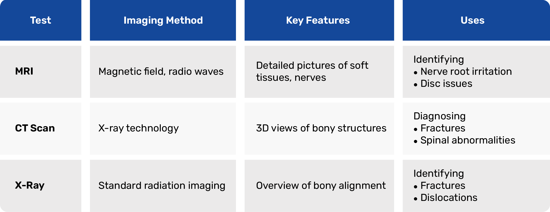
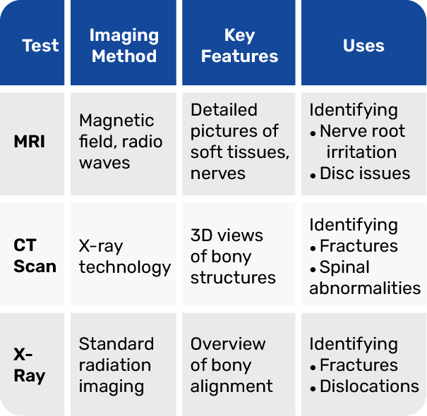
While MRIs excel in visualising soft tissues, CT scans shine in detecting minor fractures. X-rays serve as a preliminary imaging test for simpler cases.
The choice between imaging tests depends on what the doctor finds during your check-up. MRI provides excellent detail of the spinal canal and lumbar spine, making it a suitable choice when nerve problems or structural abnormalities are suspected. CT is better if you have a trauma or a possible bone break, because it can see bones very well. X-rays are often the first step in basic checks and the starting point for investigating the issue.
When you match your back pain symptoms to what these tests can do, it helps clinicians choose the best test for you. For most cases of low back pain, imaging techniques like MRI or CT are only necessary if you have red flags that could indicate a more serious condition.
Magnetic resonance imaging (MRI) is a valuable tool for evaluating conditions in your back. Still, it is not always the first diagnostic test doctors use when you have low back pain. They will look for signs of serious trouble, like nerve damage, broken bones, or tumors. If you have any of these, your doctor may recommend an MRI. Clinicians may also use magnetic resonance imaging if you have been using other treatments for a while, but you are still in pain.
However, if you have acute low back pain without any worrying symptoms, imaging like an MRI is usually not needed. So, when is magnetic resonance imaging really necessary, and when can it be skipped? Let’s take a closer look.
Some signs show there could be nerve damage or a serious problem, and you need to get an MRI right away. Watch for the following:
If you experience any of these signs, you should consult a doctor immediately for medical attention, an MRI, or other imaging tests. This is because back pain, numbness, and similar symptoms may indicate nerve damage that requires prompt care.
MRI tests are usually not needed for most cases of acute low back pain. Here are some times you can avoid imaging:
It is often better to monitor back pain and take the right steps instead of jumping to imaging. This can help lessen unnecessary worry and promote better health.
While imaging tests like MRI and CT scans can help doctors identify significant health issues, using them too frequently may pose risks to individuals. CT scans use radiation, and sometimes contrast dyes are given. This can cause people to worry more or lead to health problems. Also, MRIs can show things that are not important, and this may lead to more tests or treatments that are not needed and cost a lot. If we only use imaging techniques like MRI and CT scans when truly necessary, we achieve better results. It is essential to understand the limitations of these tests so that your safety and well-being come first.
MRI scans are generally safe for most people, but it’s essential to be aware of certain risks. If you have loose metal objects in your body or some types of implants, the strong magnetic field can cause problems. Some people who get a gadolinium shot before imaging may have a rare allergic reaction or problems with their kidneys. Pregnant women are usually told not to get this test. It is important to work with your health care provider to make sure the right screening and preparation are done. This will help lower most risks.
Too much imaging can lead to confusion in interpreting results. Some things observed in the images, such as changes associated with aging, may not be related to the symptoms the patient experiences. This can lead to unnecessary worry or additional treatments that are not necessary. When people rely too heavily on imaging, it can also label patients in a way that prolongs their negative feelings and hinders their recovery. That is why clinicians want to use imaging only when needed for diagnostic triage, and check on progress by using as few scans as possible. This helps achieve better results for patients and prevents them from unnecessary worry.
Treatment for back pain does not always require starting with imaging, such as MRI or CT scans. Many people with acute low back pain get help from simple approaches such as physical therapy and medicine. Clinicians recommend checking in on patients frequently to monitor for changes or improvements. You do not have to use expensive MRI or CT tests to do this. These practical ways help people with low back pain recover faster and save money.
Conservative care can often help people get better from acute low back pain. Most of the time, you do not need imaging for this. Some common ways to treat low back pain are:
When you make changes to your everyday habits in conjunction with these treatments, you make it easier for your back to heal on its own. These steps support a more natural approach to recovering from back pain or acute low back pain.
Tracking your improvement without imaging involves monitoring whether your pain is decreasing and assessing your ability to do more each day. Your healthcare team can recommend that you see a specialist if you do not improve with basic treatments over time. With regular visits, it is easier to spot if symptoms worsen or if any red flags appear. Most people simply need easy ways to track their progress and some good advice from their doctors. This can help you avoid excessive scanning and allow your body to heal as it should.
In short, knowing when you need an MRI or other scans for low back pain helps you make the best choices for your health. Some people think images always help, but it is good to remember both the benefits and any risks or limits of scans. Most of the time, you and your doctor should look at your medical history, do a physical exam, and pay close attention to your symptoms. This way, you often do not need a scan right away for back pain or low back pain.
It is also smart to first try basic treatments and see how you progress over time. Managing your low back pain starts with taking these steps and doesn’t need imaging early on. If you are unsure about what to do, you can consult with a healthcare professional. They can help you find the treatment that is best for you and your low back. Remember, prioritizing your health and checking in when something doesn’t seem right can help you more than you think.
No, you do not always need an MRI for back pain or low back pain. Doctors usually use imaging, like an MRI, only if they see red flags. These red flags may indicate nerve damage or a more severe underlying health issue, such as an undiagnosed pathology. Most of the time, your doctor can find the cause of your low back pain with a simple exam and a look at your medical history. Usually, there is no need for any imaging if there are no red flags.
No, MRI scans do not identify all potential causes of back pain or low back pain. They are suitable for identifying problems with your spine and other internal back issues. However, some conditions, such as nerve damage, may not be visible in MRI images. To identify these issues, the doctor will conduct a clinical examination and review the imaging results together. This helps them know what is causing low back pain.
Most people can have an MRI safely. However, some individuals may be at risk if they have metal in their body, certain implants, or kidney problems. The contrast dye gadolinium should not be used in certain individuals. You should discuss this with your doctor before undergoing the MRI.
Before the test, please remove all metal items, such as jewelry or hearing aids. This is important so the magnetic field works right. If the doctor plans to use contrast material, inform your doctor about any kidney problems or allergies you may have before the test.
Discuss your medical history with your doctor before undergoing the imaging test. Tell them if you have metal implants or any other reasons that might keep you from having an MRI. Explain why you need the MRI. Check with your clinician to determine if the imaging test is necessary due to any red flags or is part of your current treatment regimen.
Pioneering Interventional Pain Management
Delhi | Faridabad | Gurugram | Ghaziabad | Noida | Lucknow | Jaipur | Mumbai
© 2025 Nivaan. All Rights. Reserved


")


